Wednesday, January 16, 2013

Discount plans: Packing in the penny-pinching patients


Discount plans: Packing in the penny-pinching patients

September 17, 2007 -- "Buy one, get one free!" "On sale!" "Half-price with this coupon!" Such come-ons can be effective in luring customers to supermarkets, drug stores, and restaurants. Now dentists can tap similar marketing techniques via dental discount plans.
The plans may pack your office with new patients. But as an October 2006 article in the Academy of General Dentistry's AGD Impact newsletter reports, such schemes come with pitfalls as well as promise.
The basic mechanism of dental discount plans is fairly simple. Patients pay an annual fee in the neighborhood of $70 or $80 and receive a list of dentists who are willing to offer procedures at a discount, typically 20 percent to 30 percent.
The patients are happy because they merely present a card to a dentist in the plan and presto, they save money. The savings increase the more procedures the patient has done.
Dentists are happy because patients who would have gone to a competitor--or not seen a dentist at all--are coming to their offices.
Not all plans are created equal. Patients may be particularly reluctant to sign up for discount plans with a small list of providers, the article notes.
Also, problems can arise when a dentist or patient misunderstands how the scheme works. "If the patient is under the impression this is insurance, that's a problem," Tom Limoli, an Atlanta-based insurance consultant told DrBicuspid.com. “It's not insurance. It's marketing on behalf of the dentist." The confusion is not uncommon, since some companies sell both insurance and discount plan, and some vendors blur the distinction in their advertising. If nothing else, patients that have insurance and a discount plan may have problems sorting the two out.
Because of such complaints, state regulators are starting to get involved, forcing vendors to be more forthcoming.
Another drawback for dentists can be confusing contracts. According to the AGD Impact article, some contracts force the dentist to honor discounts offered by the carrier's affiliates or subsidiaries, compelling the dentist to offer different sets of discounts. When carriers merge with other companies, the terms of the plan can also change on short notice. Typically, says the report, dentists can extricate themselves only by canceling all plans and giving the carrier and affected patients two to three months notice.
The most telling problem for dentists, says Limoli, is that plan patients pay less. Since there is no insurance company making up the difference, dentists make less money off these patients. Nor are plan patients likely to spring for the sort of elective procedures that can fatten a dentist's bottom line. "[Plans do] bring in patients, but it's a type of patient that is very thrifty," Limoli said.
To make up for the lost income, says Limoli, dentists must trim their administrative costs. Since there is no paperwork to file with an insurance company, a dental discount plan can save office staff time. The key is requiring plan patients to pay upfront. If they can't afford to pay on the spot, dentists should ask them to use credit cards or borrow money from a third party, Limoli advises.
The AGD article stops short of giving discount plans a thumbs up or thumbs down. But Limoli, while cautioning against the pitfalls inherent in these plans, thinks they can work to everyone’s advantage because they reach a population that typically avoids dentists. "Many people don't seek dental care because it's too expensive," he says. "Dentistry has to be more available to more people."

LCT catches long fractures -- in the lab


LCT catches long fractures -- in the lab

September 19, 2007 -- A kernel of popcorn that went unpopped. A piece of ice like an iceberg. Grueling nighttime grinding. Who knows what started a fissure down the length of that tooth?
As the pain mounts, so does the frustration, because longitudinal tooth fractures -- which extend through the long axis of the tooth and expand with time -- are often invisible to the naked eye and difficult to detect with conventional radiology.
Such teeth can rarely be saved, so dentists and patients would both benefit from discovering a fracture right away. "It would prevent a lot of unnecessary treatment and suffering," says André Moll, DDS, MS, PhD, an Oral and Maxillofacial Radiologist and Assistant Professor of Diagnostic Science and General Dentistry at the University of North Carolina, Chapel Hill.
That’s why Moll and his colleagues were eager to try out local computed tomography (LCT) as a method of detecting these problem cracks. This new technology, which is a form of cone beam CT, produces high resolution 3D images of a small area of the jaws
As they reported in the June 2007 Oral Surgery,Oral Medicine, Oral Pathology, Oral Radiology, and Endodontology, the method works well -- at least when applied to extracted teeth under laboratory conditions.
In their experiment, the researchers anchored 30 whole teeth in acrylic, one by one, to prevent the roots from splitting, then coated them with wax for easy removal. Then they placed a screwdriver-style in the canals and gently tapped, fracturing the teeth from within the root. These 30 split teeth were matched to 30 whole teeth that were used as controls.
To simulate conditions within the mouth, they placed the teeth in an empty socket of an otherwise dentate mandible, and before scanning they surrounded the mandible with boxing wax, which simulated soft tissue. Silicone dental wax served as a surrogate for trabecular bone, and the researchers mixed in soy grains to simulate marrow spaces.
Then the scanning began. The investigators used a conventional cone-beam dental x-ray source, the Planmeca Prostyle Intra, and a Schick CDR detector, which has high-resolution digital capacity. The mandible sat on rotator  and the detector matrix was aligned with the rotational axis of the teeth.
The source sat 65 cm from the tooth, and the detector sat 12.5 cm away. As each tooth was scanned it was placed in the center of rotation, with the long axis of the tooth parallel to the rotation axis. This allowed the system to take 180 images with 1 degree of separation in a 180-degree arc. This created 3D images, which the investigators hoped would offer an advantage over conventional 2D x-rays. Conventional x-rays usually can't show fractures unless the x-ray beam is parallel to the plane of the facture.
A filtered back projection reconstruction algorithm allowed the investigators to generate multiple axial slices with a thickness of 0.2 mm. They removed the top slices showing the crown to prevent observers from judging the fractures based on the status of the crowns, which were damaged in producing the fractures.
For comparison purposes, the researchers took standard 2D x-rays of each tooth using an Eastman Kodak RVG 6000 sensor. To likewise prevent the observers from being clued in, they masked the crowns with an opaque overlay.
Finally, the investigators put their images to the test, showing them to four radiologists, one radiology student, four endodontic residents, and a periodontist. Using three side views (orthogonal planes to view the image volume),  the observers were asked to rate the probability on a scale of 1 to 5 that each tooth had a fracture.
The results suggest that LCT is significantly more accurate than traditional radiography. The investigators tested the observers’ ratings of the teeth by plotting their "false positive" responses against their "true positive" responses and measuring the area under the curve (a receiver operating characteristic curve). The observers scored an average of 0.91 with LCT vs. 0.70 with conventional radiography. That's equivalent to a 91 percent versus 70 percent accuracy rate, had the observers given simpler "yes or no" answers to the question of whether each tooth was fractured (a two-alternative forced choice comparison), Moll said.
The UNC researchers cautioned that the results don’t prove the new technology will work in a real-world situation with a human mouth. Real-life fractures may not resemble a tooth split with a screwdriver. Nor was there real bone or ligament surrounding the teeth in this experiment. Likewise, in actual practice, the teeth with longitudinal fractures dentists encounter may already be restored in some way, often with posts.
These differences between laboratory conditions and the real world may explain why the observers were able to identify so many of the fractured teeth using conventional radiography -- a finding that somewhat surprised the investigators.
Engineers must resolve another problem before the technology is truly practical: creating a 3D image from 180 images meant using 180 times as much radiation as in the standard x-ray, an unacceptably high dose.
"We've done some research since then to see if we could bring the dosage down," said Moll. "We were successful, but it's really a problem we need to work with our industry partners to solve."
Overall, he said, LCT has significant potential to help solve a problem that has bedeviled dentists since the profession began.

Copyright © 2007 DrBicuspid.com

Dental assistants at risk of lung problems


Dental assistants at risk of lung problems

Methacrylates are used in dental filling materials and bonding agents, like those used to cement porcelain veneers, crowns, and orthodontic brackets. Dental assistants are exposed to airborne methacrylate particles when mixing these materials or during placement or removal of dental restorations.
In the new study, researchers found that among 799 Finnish dental assistants, those with greater methacrylate exposure had higher risks of developing asthma or respiratory problems like chronic nasal symptoms, hoarseness, and breathing difficulty.
"The results suggest that exposure to methacrylates poses an important occupational hazard for dental assistants," the study authors report in the journal Allergy.
"The risks to respiratory health are related to inhaling these substances," lead author Dr. Maritta S. Jaakkola, of the University of Birmingham in the UK, told Reuters Health.
Probably the most important protective measure is for dentists to install exhaust systems in areas where assistants work with methacrylates, Jaakkola said.
The findings are based on questionnaire responses from 799 female dental assistants. The researchers asked the women how often they performed tasks like mixing dental fillings and sealings, and whether they'd been diagnosed with asthma or frequently suffered respiratory symptoms -- like a stuffy nose, cough or breathlessness.
Overall, the study found, women who'd been exposed to methacrylates every day for the past three months were nearly three times more likely than less-exposed dental assistants to report adult-onset asthma. They also showed higher risks of nasal symptoms and work-related coughing.
The risk of respiratory symptoms appeared to grow the longer women had been on the job, and those who'd suffered allergies as children seemed particularly susceptible.
In general, dental assistants who reported daily exposure to methacrylates for more than 10 years had higher risks of hoarseness, breathlessness, and wheezing. Among assistants with a history of childhood allergies, those who reported daily methacrylate exposure had a four-fold increased risk of adult-onset asthma, and a two-fold higher rate of nasal symptoms.
Besides exhaust systems to clear the air, gloves also offer dental assistants protection from methacrylates, Jaakkola noted. The substances can cause skin reactions, she explained, and it's also possible that sensitization to methacrylates through skin contact makes some people more susceptible to suffering respiratory effects as well.
Allergy, June 2007.
Copyright © 2007 Reuters Limited. All rights reserved. Republication or redistribution of Reuters content, including by framing or similar means, is expressly prohibited without the prior written consent of Reuters. Reuters shall not be liable for any errors or delays in the content, or for any actions taken in reliance thereon. Reuters and the Reuters sphere logo are registered trademarks and trademarks of the Reuters group of companies around the world.

Fentanyl provides effective supplemental analgesia in dental surgery for pulpitis


Fentanyl provides effective supplemental analgesia in dental surgery for pulpitis

Standard pain relief in dental debridement relies on interligamentary injections of mepivacaine and epinephrine. However, local analgesia is often inadequate because the acid environment created by the inflammation prevents uptake of the anesthetic, principal investigator Dr. Eman A. Elsharrawy and colleagues at October 6 University in Cairo point out.
Forty patients with irreversible pulpitis received an initial interligamentary injection of 1.8 ml of 2% mepivacaine plus epinephrine 1:200,000, which is standard therapy for dental debridement.
Patients were then randomized to receive supplemental fentanyl, 0.4 ml in a 0.05 mg/ml solution added to a supplementary course of 0.4 ml 2% mepivacaine with epinephrine 1:200,000 or to supplemental local anesthesia alone. The drugs were given in two divided doses, with 0.2 ml given on the mesial side of the tooth and 2 ml given distally.
Fentanyl provided significantly greater pain relief during various stages of surgery, including no pain on pulp extraction, than the standard approach with mepivacaine plus epinephrine. Patients randomized to local anesthesia only experienced more pain at all stages of surgery.
The findings "confirm the importance of peripheral opioid actions in inflammatory pain and suggest the value of this approach for the clinic," Dr. Elsharrawy and colleagues write. In contrast with its effect on local anesthetic agents, an acid environment produces "enhanced opioid effects."
J Pain Symptom Manage 2007;33:203-207.
Copyright © 2007 Reuters Limited. All rights reserved. Republication or redistribution of Reuters content, including by framing or similar means, is expressly prohibited without the prior written consent of Reuters. Reuters shall not be liable for any errors or delays in the content, or for any actions taken in reliance thereon. Reuters and the Reuters sphere logo are registered trademarks and trademarks of the Reuters group of companies around the world.

Defining a role for MRI in TMJ disorders


Defining a role for MRI in TMJ disorders

July 19, 2007 -- Chances are that dentists won't have to worry about siting a magnet in their offices in the near future, but can the specialty use MRI to its advantage? Yes, if the structure in question is the temporomandibular joint (TMJ). "Particularly, we look at this disk in the TMJ because (MRI) is the only way we can see the disk. MRI gives you exquisite soft-tissue imaging that you can't get with other imaging," said Dr. Edwin Parks in a talk at the 2006 California Dental Association meeting in San Francisco.

Indeed, over the latter half of the 20th century, MRI has replaced arthrography and arthrotomography as the go-to modality for visualizing the soft tissues of the TMJ, as well as the overall integrity of the structures.
Specifically, dentists need to look at the relationship of the articular disk to the head of the condyle and the head of the glenoid fossa, added Parks, who is director of dental radiology at the Indiana University School of Dentistry in Indianapolis.
Parks recommended that dentists consider MRI for assessing soft-tissue alterations in the TMJ, inflammation, disk displacement, and joint effusion. Researchers in Japan did exactly that, using MRI to pinpoint TMJ disorders and then comparing them to discrepancies of the glenoid fossa.
But not everyone is sold on TMJ MRI. A recent paper analyzed published research on TMJ MRI, questioning whether enough evidence existed to make MRI part of the routine clinical protocol.
RGF and TMJ on MRI
To determine if thickness of the roof of the glenoid fossa (RGF) correlated with TMJ disorders found on MRI, Japanese dentists studied 87 patients with TMJ disorder symptoms who were referred for MRI.
"Little is known about changes in the thickness of the RGF under different normal and pathological conditions. It might be that an increase in joint thickness is a natural response of the body in order to maintain proper joint function," wrote Dr. Kazuya Honda, Ph.D., from Nihon University School of Dentistry in Tokyo. "The present report ... measured the minimum thickness of the RGF in relation to MRI observations of TMJ structure" Dentomaxillofacial Radiology), September 2006, Vol. 35:5, pp. 357-364).
The MR protocol consisted of T1- and T2-weighted images, in the sagittal and coronal planes, in a closed mouth. T2*-weighted images were also obtained in the sagittal plane with a closed mouth and the mouth open to maximum. A 1-tesla scanner was used (Magnetom Harmony, Siemens Medical Solutions, Malvern, PA). Three-dimensional conebeam CT images (3DX Accuitomo, Morita, Kyoto, Japan) were also acquired to measure the thickness of the RGF at its thinnest point.
According to the authors, the "results showed that there was no significant difference in the minimum thickness of the RGF between normal joints and those classified as ADWR (anterior disk displacement with reduction) or ADWOR (anterior disk displacement without reduction)."
More specifically, there were 70 normal joints, 53 joints with ADWR, and 51 joints with ADWOR. The average minimum thickness of the RDF was 0.85 mm in normal joints, 0.90 mm in ADWR joints, and 0.93 mm in ADWOR joints.
Based on additional analysis, there were 21 joints with osteoarthritis (OA), 153 without OA, 61 with joint effusion, and 113 without joint effusion. The RGF thickness in the OA group was 0.99 mm versus 0.87 mm in the group without OA. RGF thickness in 33 joints with disk deformation was 0.87 mm compared to 0.89 in 141 joints without disk deformation.
However, there was a significant RGF thickness between the groups with joint effusion (0.97 mm) and those without joint effusion (0.84 mm), the authors stated, indicating that RGF thickness is influenced by joint effusion.
While the minimum thickness of the RGF in TMJ did not correlate with disk position or configuration, the increased RGF thickness in OA and joint effusion might indicate a relationship between that thickness and pain, which could be relevant clinical information for treatment planning, the authors stated.
MR results equal better outcome?
In another study, a group from Sweden performed a systematic literature review of the efficacy of MRI in TMJ disorders. While their results did not dismiss the use of MRI in these patients, they did point out the current paucity of high-quality studies on MRI in TMJ.
"Although MRI of the TMJ has now been used for many years, its diagnostic and therapeutic efficacy -- the value of MRI for supporting clinicians in their diagnoses and treatment decisions -- remains unclear," wrote Dr. Napat Limchaichana and colleagues from the faculty of odontology at Malmö University in Malmö, Sweden (Oral Surgery, Oral Medicine, Oral Pathology, Oral Radiology, and Endodontology, October 2006, Vol. 102:14, pp. 521-536).
For this study, the authors mined the PubMed database from January 1988 to July 2005 and found 494 titles. Two reviewers looked these titles over, assessing the level of evidence in favor of TMJ MRI as high, moderate, or low. They also graded the studies for diagnostic efficacy as strong, moderately strong, limited, or insufficient. Ultimately, 22 papers met the study criteria for level of evidence and diagnostic efficacy.
Of those 22 articles, none offered a high level of evidence on the value of MRI in TMJ disorders, according to the authors. Only a dozen offered moderate evidence and 10 had low levels of evidence. More specifically, the studies did not demonstrate a consistent likelihood that MRI could diagnose TMJ disk position.
In terms of TMJ disk configuration, studies showed that the specificity (50%) and positive predictive value (56%) of MR sagittal images were low for identifying biconcave versus deformed disks. Finally, MR turned in only moderate results for identifying osseous changes of the TMJ and bone marrow changes.
"We ... conclude that evidence for observer performance is limited, and evidence for diagnostic efficacy ... is insufficient," the authors wrote. "However, insufficient scientific evidence for a method's efficacy does not necessarily imply that the method is ineffective or should not be used."
Instead, they called for additional studies based on validated methodology for determining diagnostic and therapeutic efficacy. In the 22 studies they reviewed, there was too much variation between diagnostic criteria, the authors stated.
"An ideal appraisal should check observer reproducibility.... Confidence intervals for sensitivities, specificities, and predictive values should also be determined.... At present, it is (impossible) to draw any conclusion about when the results of an MRI examination will result in better treatment outcome for the (TMJ disorder) patients," they concluded.

Chronic periodontitis linked to tongue cancer


Chronic periodontitis linked to tongue cancer

"We expected to see an association, given the results of earlier studies linking chronic infections and inflammation to cancer risk in other organs," lead author Dr. Mine Terzal, from the University at Buffalo School of Dental Medicine in New York, said in a statement. However, "we didn't expect to see such a clear association with a relatively small sample size."
The study involved 51 white men with newly diagnosed squamous cell cancer of the tongue and 54 cancer-free controls who were seen during the same period. The study excluded people younger than 21 years and those who lacked teeth, were immunocompromised, or had a history of any malignancy. Alveolar bone loss on radiography was used to assess periodontitis.
The researchers' report their findings in the May issue of the Archives of Otolaryngology: Head and Neck Surgery.
On multivariate analysis, each millimeter reduction in alveolar bone was associated with a 5.23-fold rise in the risk of tongue cancer, the report indicates.
"Periodontitis is a chronic disease that progresses very slowly," Dr. Terzal noted. "Seeing alveolar bone loss on x-rays indicates the infection has existed for decades, making it clear that periodontitis preceded the cancer diagnosis, and not vice-versa."
The authors conclude that larger studies are needed to confirm these findings and to better control for lifetime tobacco exposure.
Copyright © 2007 Reuters Limited. All rights reserved. Republication or redistribution of Reuters content, including by framing or similar means, is expressly prohibited without the prior written consent of Reuters. Reuters shall not be liable for any errors or delays in the content, or for any actions taken in reliance thereon. Reuters and the Reuters sphere logo are registered trademarks and trademarks of the Reuters group of companies around the world.

Fake dentist has 29-year career in Malaysia


Fake dentist has 29-year career in Malaysia

The impostor's closest brush with the dental profession was during the years 1962 to 1978, when he assisted an army dentist by carrying his bag on visits to plantation workers' homes, the New Straits Times reported Wednesday.
"I watched the doctor diagnose and treat problems with teeth," the paper quoted the unidentified man as saying when officials raided his home this week.
"I also saw how he would extract teeth and make models and measurements for dentures."
The 63-year-old man, who convinced his neighbors he was a retired army dentist after being told he was too old to work as a dentist's assistant, charged 20 ringgit ($5.76) for extractions and 130 ringgit for dentures, the paper said. ($1 = 3.475 Malaysian ringgit)
Health officials seized antibiotics, painkillers, syringes and bottles of Chinese medicine during the raid, but they needed more than six men to carry off his 1940s-era examining chair, tossed away by the Malaysian army in 1978. The paper said the raid occurred after a tip-off.
The man has been arrested for illegally practicing dentistry and will face charges under Malaysia's private healthcare facilities and services act. He could face a fine of up to 30,000 ringgit or a six-year jail term, or both.
He did provide one unique service, however. "I also make house visits," he told the paper.
Copyright © 2007 Reuters Limited. All rights reserved. Republication or redistribution of Reuters content, including by framing or similar means, is expressly prohibited without the prior written consent of Reuters. Reuters shall not be liable for any errors or delays in the content, or for any actions taken in reliance thereon. Reuters and the Reuters sphere logo are registered trademarks and trademarks of the Reuters group of companies around the world.

Tooth loss = higher heart disease risk?


Tooth loss = higher heart disease risk?

The findings, reported in the journal Heart, add to evidence linking oral health to heart health. A number of studies have suggested that gum disease may contribute to heart disease over time -- though it's still not clear that there is a cause-and-effect relationship.
This latest study involved more than 12,000 UK adults who were followed from college onward, for up to 57 years. Researchers found that those with a large number of missing teeth in young adulthood -- nine or more -- were one-third more likely to die of heart disease than their peers with fewer than five missing teeth.
The link remained when the researchers considered factors such as socioeconomic background and smoking, which harms both the teeth and gums and the heart.
Tooth loss is an indicator of poor oral health. Scientists speculate that the bacteria in the mouth that cause cavities and gum disease may enter the bloodstream and either damage the blood vessel lining directly or trigger inflammation in the body that then contributes to heart disease.
The current findings do not prove that this is the case, according to the study authors, led by Dr. Yu-Kang Tu of the University of Leeds. But they do support a relationship between tooth loss and cardiovascular disease, Tu told Reuters Health. Unlike most other studies in this area, the researcher noted, this one looked at oral health early in life rather than in old age.
"Our study adds to the evidence that chronic infection -- oral or elsewhere in the (body) -- may increase the risk of cardiovascular diseases," Tu said.
The findings are based on 12,631 men and women who had medical and dental exams as college students in the 1940s through 1960s. They were then traced through the UK National Health Service until 2005, during which time 1,432 died.
Overall, men and women with the most severe tooth loss as college students were 35 percent more likely to have died from heart disease than those with four or fewer missing teeth.
It's too soon to say that good oral hygiene will lower anyone's risk of heart disease, according to Tu -- particularly since this risk depends on multiple lifestyle and genetic factors. But, Tu added, it also won't hurt people to take better care of their teeth.
Heart, September 2007.
Copyright © 2007 Reuters Limited. All rights reserved. Republication or redistribution of Reuters content, including by framing or similar means, is expressly prohibited without the prior written consent of Reuters. Reuters shall not be liable for any errors or delays in the content, or for any actions taken in reliance thereon. Reuters and the Reuters sphere logo are registered trademarks and trademarks of the Reuters group of companies around the world.

New evidence restores amalgam's image


New evidence restores amalgam's image

September 11, 2007 -- Do a Google search for "amalgam" and "danger" and you get over 460,000 hits. Small surprise. Health advocates rail against the danger of "silver" mercury (amalgam) fillings. Some even claim that removing existing amalgam restorations will radically improve patients' health. Meanwhile, amalgam boosters staunchly defend the alloy.
But in the decades that amalgam has been on trial, neither the prosecution nor the defense has found enough evidence to win its case. That's why two long-term studies reported last year in the Journal of the American Medical Association (JAMA), attracted wide-spread attention: both suggested that mercury fillings don't affect children's mental or physical health.
Predictably, amalgam defenders such as the American Dental Association hailed these results. And just as predictably, amalgam critics rushed to poke holes. Now both studies are back in the news, with new articles--including one in the September issue of the Journal of the American Dental Association (JADA)--in which the researchers address some of the major criticisms. The bottom line: the results still stand.
“There appear to be no detectable adverse neuropsychological outcomes in children that are attributable to the use of amalgam dental restorations,” concluded the researchers of The New England Children’s Amalgam Trial in the most recent article.
Researchers from both studies also gave the pro amalgam camp further ammo by publishing evidence that amalgam fillings last longer than composite fillings.
Exposed, yes. But harmed?
Dentists have used amalgam to repair teeth for 150 years. But amalgam contains 40% to 50% mercury by weight, and it emits minute amounts of mercury vapor under the pressure of ordinary chewing.
Both 2006 studies compared the health of a group of children with amalgam fillings to the health of a group with composite fillings over several years.
For the New England Children’s Amalgam Trial, researchers at Harvard University, four other U.S. institutions, and Goteberg University in Sweden, randomly divided 534 Massachusetts children into two groups of 267 each. The children were 6 to 10 years old at the start of the study, and had at least two caries in posterior teeth. Over the next five years, the children had a mean of 15 restorations.

After five years of getting their cavities filled, the children who received amalgam restorations had nearly 50% more mercury in their urine as compared to the group getting composite fillings (0.9 µg/g vs. 0.6 µg/g). But based on the results of a battery of neurocognitive tests evaluating intelligence, memory, and visuo-motor skills, both groups did equally well. Nor did the amalgam children’s kidneys appear to be affected by the mercury. There was no significant difference in urinary albumin between the amalgam kids and the composite kids over the five years.
Researchers for the Casa Pia Study of the Health Effects of Dental Amalgams in Children from the University of Washington, Seattle; the Battelle Centers for Public Health Research and Evaluation in Seattle; and the Lisbon-based Universidade de Lisboa and the Universidade Catolica Portuguesa; randomly divided 507 Lisbon children into a group of 253 who received amalgam restorations and a group of 254 who received composite restorations. The children ranged in age from 8 to 10 years and each had at least one carious lesion on a posterior tooth.
After seven years, the children had a mean of 20 restorations. As in the New England study, the amalgam children had a higher creatinine-adjusted level of mercury in their urine after two years (3.2 µg/g vs 2.7 µg/g), but that difference shrunk between the two groups over the seven years. And there were no statistically significant differences in memory, attention, and visual motor function or nerve conduction velocities at any point during the seven years.
Both groups of researchers argue that their studies should make dentists more comfortable using amalgam. But in an editorial accompanying the two JAMA reports, Herbert L. Needleman, M.D., a Pittsburgh University pediatric psychiatrist pointed out several shortcomings:

  • The damage from mercury exposure could accumulate over decades. The longest-term follow-up in these two studies was seven years.
  • The studies were not large enough to detect subtle, but potentially important, brain damage.
  • There is evidence that genetic differences make a portion of the population highly vulnerable to mercury. Neither study focused on the effects in patients with these genetic differences.
  • The studies didn’t examine whether those children with the most mercury in their urine experienced neurological effects; it simply lumped all the children together.

Upon further inspection…
In response, the New England researchers further analyzed their data and published two follow-up reports, the first in the March 2007 Journal of Environmental Health Perspectives and the second in JADA this month. Both articles compared the amount of mercury in children’s hair and urine and the number of restorations per child to the detailed children’s scores on an extensive battery of neurological tests.
Once again, the New England researchers said the results looked good for amalgam. Even when they ranked the children in order of least-to-most mercury exposure, they found no evidence of neurological damage.
And zooming in on the minutia of the test scores didn't reveal any problems, either. In only two out of 32 of these tests did the researchers find significant differences between the amalgam kids and the composite kids (the Number Letter Memory subtest of the Wide Range Assessment of Memory and Learning and the 4-year change in the time required to complete Part B of the Trail-Making Test). And these correlations didn’t implicate amalgam. In fact, the children with amalgam fillings scored higher than the children with composite fillings on both tests.
The researchers also did not find any subgroup of children who suffered any effects from mercury exposure. They admitted that their study was too small and short term to completely rule out the possibility that amalgam fillings cause brain damage, but they argued that dentists can be reasonably confident in using the material until further research proves otherwise.
In fact, in a separate June 2007 article in JADA, the New England researchers said there’s a good reason to prefer amalgam--it lasts longer than composite fillings. In primary teeth, 3.0 percent of composite fillings needed replacement due to caries vs 0.5 percent of amalgam fillings over the five years.
The Casa Pia researchers reported similar findings about the durability of amalgam in the same issue of JADA. In short, say the Casa Pia authors, "amalgam restorations performed better than composite restorations."
Funding Facts:
Both studies received funding from the National Institutes of Health. The New England study also got money from the New England Research Institutes. Only two researchers--from the Lisbon studies--made financial disclosures. One received a small grant from an amalgam restorations company to travel to Washington state. The other consults for a company that makes both composite and amalgam restorations.
Sources:
Bellinger, C et al., "Dental Amalgam Restorations and Children’s Neuropsychological Function: The New England Children’s Amalgam Trial" Environmental Health Perspectives 115:440–446, March 2007
Bellinger, C et al., "Neuropsychological and Renal Effects of Dental Amalgam in Children: A Randomized Clinical Trial" Journal of the American Medical Association, Vol 295, No. 15, April 19, 2006.
Bellinger, C. et al., "A dose-effect analysis of children’s exposure to dental amalgam and neuropsychological function: The New England Children’s Amalgam Trial" Journal of the American Dental Association, 138(9):1210-6, Sept 2007
Bernardo, M. et al., "Survival and Reasons for Failure of Amalgam Versus Composite Posterior Restorations Placed in a Randomized Clinical Trial," Journal of the American Dental Association,138(6):775-8, June 2007
Rouen, T. et al., "Neurobehavioral Effects of Dental Amalgam in Children: A Randomized Clinical Trial," JAMA, Vol 295, No. 15 April 19, 2006

Dental tourism on the rise?


Dental tourism on the rise?

September 13, 2007 -- A growing number of U.K., American, and Japanese citizens are including expensive dental procedures in their vacation plans this year. According to Ireland-based RevaHealth.com, a medical tourism search site, nearly a million consumers go abroad every year to have their teeth treated. According to Reva, consumers save as much as 63% on dental procedures performed in Mexico, Hungary, Thailand, and other countries with much lower costs of living and favorable exchange rates.
Dental tourism is part of the larger medical tourism trend that is sweeping developed nations as medical costs continue to skyrocket and waiting lists get longer and longer. From hip replacements and bypass surgeries to breast implants and tooth implants, hospitals in Thailand, India, Mexico, and the former Eastern Bloc offer cheaper, timely, and according to advocates, high-quality medical care. Patients who are willing to travel halfway around the world for an exotic vacation-cum-medical procedure can potentially save a bundle.
According to Reva, medical tourism is a $20 billion industry and growing steadily, with 500,000 Americans alone last year seeking medical treatment outside the U.S. Although there are no hard numbers, dental tourism seems to be going along for the ride. According to Reva, over 40,000 Americans went to Mexico for dental treatment last year, saving an estimated 75% compared to typical fees in the U.S.
"U.S. dental fees are getting higher and higher and that's why dental tourism has grown in the past three or four years," says Helga Vanhorn, who runs a medical tourism service in Nevada with her husband. Most of her clients have little or no insurance, and with a $1000 crown costing only $300 in Hungary, the lure is tempting. How much can patients save? Reva's site sports a "Dental saving calculator." Simply check the procedures you want (such as implants, whitening, dentures), click a button, and the calculator shows you how much you'd spend in the U.S. -- and how much you'd supposedly save going to Costa Rica, Mexico, and other countries.

Is dental work cheaper outside the U.S.? According to Reva's calculator, you can save a bundle.
Is dental tourism taking a big chunk out of American dentists' bottom line? "There's no indication of that whatsoever," says Dr. Edmond Hewlett, Consumer Advisor for the ADA, but he admits that dental tourism is seemingly on the rise.
What about patient safety? Again, there are no hard numbers, but the British Academy of Cosmetic Dentistry expressed doubts about overseas dental care in a November 2006 press release. "Dentists ... are seeing a surge in cases of botched treatment: patients coming back to the U.K. with badly fitted crowns and mismatched veneers, and worst of all, nerves removed for no apparent reason," says the release.
Gayle Mathe, Manager of Policy Development at the California Dental Association, considers the lack of follow-up care to be a key disadvantage of dental tourism. As the ADA's Hewlett points out, dental tourists are often seeking complex work. Patients should confirm the credentials of any overseas dentist before taking the trip, and carefully evaluate the host country's safety/hygiene regulations and legal safeguards.

Calcium during breast-feeding may guard mom's teeth


Calcium during breast-feeding may guard mom's teeth

August 14, 2007 -- NEW YORK (Reuters Health) Jan 22 Women who breast-feed may need to be careful about getting enough calcium to keep their teeth and gums healthy, new animal research suggests.
In experiments with rats, researchers found that lactating rodents were particularly susceptible to the effects of low calcium intake on the bones that support the teeth. Such bone-density loss can speed the progression of any existing gum disease.
Though the findings come from animals, they do suggest it's important for breast-feeding mothers to include enough calcium in their diets, lead researcher Dr. Kanako Shoji told Reuters Health.
Shoji and colleagues at Tohoku University Graduate School of Dentistry in Japan report the findings in the Journal of Periodontology.
During breast-feeding, a woman's calcium demands go up to meet her growing baby's needs, the researchers point out. In addition, certain hormonal changes during breast-feeding may contribute to bone-density loss.
So adequate calcium intake -- from foods like milk, cheese, and fortified cereals and juice -- may become particularly important. The recommended calcium intake for women ages 19 to 50, breast-feeding or not, is 1,000 milligrams a day.
If a woman doesn't get adequate calcium from food, Shoji noted, supplements are an alternative.
For their study, the researchers fed lactating and nonlactating rats a diet with either adequate or low calcium levels. Periodontitis (a gum disease) was induced on one side of the animals' mouth.
The researchers found that all of the animals on the low-calcium diet lost bone density on the periodontitis-affected side of the mouth. However, the extent of the loss was greater in the lactating group.
In contrast, lactating rats that were fed enough calcium showed no such effects.
This, according to the researchers, suggests that if a woman gets enough calcium, breast-feeding may not be a risk factor for dental bone loss.
Copyright © 2007 Reuters Limited. All rights reserved. Republication or redistribution of Reuters content, including by framing or similar means, is expressly prohibited without the prior written consent of Reuters. Reuters shall not be liable for any errors or delays in the content, or for any actions taken in reliance thereon. Reuters and the Reuters sphere logo are registered trademarks and trademarks of the Reuters group of companies around the world.

Mich. dentist reaches out to Allcare patients


Mich. dentist reaches out to Allcare patients

January 1, 1999 -- A dentist in Michigan is offering former patients of Allcare Dental and Dentures up to $10,000 worth of dental implant services.
Bruce Smoler, DDS, is also working to garner the support of dentists, oral surgeons, hospitals, and other medical providers nationwide to help him in this cause, he noted in a press release.
Dr. Smoler was spurred to take action after Allcare on January 1 suddenly closed down all of its 52 clinics in the U.S. -- 10 of which were in Michigan -- leaving patients without access to records, follow-up care and treatment, or much-needed teeth, even for those who prepaid thousands of dollars for their services.
"For me, the tipping point came when the chain of offices closed down, stranding thousands of patients across the country with no place to continue their care and treatment," Dr. Smoler said. "That is so completely wrong. I find it professionally reprehensible and personally disgusting."
He is urging Allcare patients to visit his website for further information and assistance.
Copyright © 2011 DrBicuspid.com

MDCT advances oral and maxillofacial surgery practice


MDCT advances oral and maxillofacial surgery practice

November 22, 2004 -- Oral and maxillofacial surgery evolved past the clay (modeling) age when high-resolution CT entered the practice. Today's MDCT-equipped center allows surgeons to assess complex trauma injuries with razor-sharp resolution.

Once the thin-section images are acquired, volume reconstruction software aids planning in difficult oral surgery procedures, allowing practitioners to straighten skewed mandibles and maxillae on the computer, getting the bite right before the surgeon lays a hand on the patient.
Not that everything has changed. Clay modeling, photography, and radiography are still the norm in most practices, and the nascent CT software market has yet to produce the perfect reconstruction application. Still, prospects seem to be improving each year as more vendors get involved, according to Dr. Jon Bradrick (DDS), who spoke last month at the American Association of Oral and Maxillofacial Surgeons (AAOMS) meeting in San Francisco.
The oral and maxillofacial surgeon shared his decades of experience in facial imaging and procedure planning with CT, dramatized with a cache of jaw-dropping case studies. Bradrick is an associate professor and director of oral and maxillofacial surgery at MetroHealth Medical Center in Cleveland, and has worked with CT since the modality's earliest days.
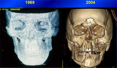
Compared to the rough, grainy images CT offered in its infancy (left), today's multislice scanners and thin-section protocols provide far more detail for maxillofacial procedure planning. All images courtesy of Dr. Jon Bradrick.
Acquisition
Today's multislice scanners allow for planning of intricate procedures in the most difficult cases, Bradrick said. The best results start with multislice scanners, thin-collimation scanning, and zero gantry tilt. Bradrick's group uses an MX8000 scanner (Phillips Medical Solutions, Andover, MA), "souped up" to be a 40-slice machine, he said.
"Here's a little trick I learned," Bradrick said. "If you think you're going to be doing some ... extreme CT reconstructions, I'll show you how to separate the patient's mandible and maxilla so you can manipulate them. Put a radiolucent object between the teeth, like a tongue blade or a piece of gauze that doesn't have a radiopaque marker in it, and just separate the teeth. That helps you when you try to segment out the mandible."
And don't administer contrast material unless you're looking for CT angiography results, he said. Prominent blood vessels can impede visualization of bone and cartilage.
When ordering the scan, be sure to specify anatomic landmarks for the radiologist -- from the supraorbital rims to the larynx, for example. Doing so helps radiologists "give me what I want, not what they think they want to give me," Bradrick said. "It has decreased our number of repeat scans."
Reconstruction software
Two major kinds of reconstruction software are in common use: applications coupled with CT scanners, such as the Philips Brilliance package, or software from standalone developers, such as Vworks 4.0 Clinic 3D by CyberMed of Seoul, Korea. Both applications were used to create the images in this article.
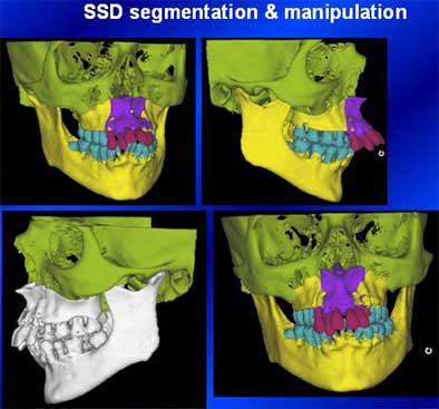
There are also two major types of reconstruction software available: traditional surface-shaded display (SSD) applications, and the newer 3D volume reconstruction products that offer complete 3D segmentation and manipulation. Reconstruction software allows the practitioner to take the CT image apart and work with it, he said, "so you can move a mandible, move a maxilla."
Both software types allow traditional 2D viewing of axial slices. Both produce minimum and maximum intensity projections, multiplanar reconstructions, and curved reconstructions, which are useful for producing dental panorexes. Both enable measurement and annotation of the anatomy for procedure planning.
However, SSD reconstruction programs get by on only about 10% of the image data, a throwback to the days of more primitive computers, Bradrick said. Offering only external topographic reconstructions, these applications apply thresholding to create a complete image from only a small portion of the CT data, resulting in poorer resolution, and more artifacts and false surfaces compared to volume 3D reconstructions, he said. And unfortunately, SSD reconstructions are the only format vendors currently use to produce stereolithographic, or 3D-printed, anatomic models.
In contrast, 3D volume reconstruction programs use 100% of the CT dataset, yielding superior image quality and the ability to depict tiny structures and fractures far better than SSD programs. The newer applications are also capable of displaying precise attenuation values and anatomic measurements, and can also produce minimum and maximum intensity projections (MIPs).
"Not a lot of people are using (volume reconstruction software)," Bradrick said. "We started using it a lot and other people started when they saw the results we were getting.... However, SSD, even with its antiquities, does do one thing for us that the newer volume reconstruction (programs) don't: segmentation and manipulation," he said.
These processes are essential to virtual surgery in the CT environment, Bradrick said. Considering the need, volume reconstruction programs that add segmentation and manipulation are certain to appear on the market eventually, Bradrick said.
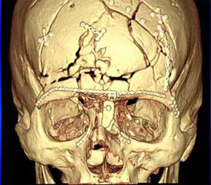
Postsurgical CT reconstruction. Image is of a young man who had suffered a serious motorcycle accident, presenting with multiple midface, frontal sinus, and frontal bone fractures. A pneumocephalus complication postsurgery required further intervention.
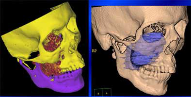
Above and below: Preteen boy presenting with facial swelling had maxillary and skull base chondrosarcoma. Presurgical planning with CT reconstructions enabled precise characterization of the lesion. At three months, surgery appears to have been successful.
Orthognathic surgery
An improper bite can give rise to a host of problems related to chewing, speech, and general oral health. The standard remedy, orthodontics, simply isn't enough when the jaw needs repositioning. Orthognathic surgery can be used to reposition the mandible, maxilla, and dentoalveolar segments to achieve facial balance and correct occlusal discrepancies.
MDCT with 3D reconstruction can replace -- and improve on -- a wide range of traditional surgical planning tools such as cephalometric and panorex radiography, dental impressions, and traditional model surgery, Bradrick said. With the aid of 3D software, malocclusions and deformities of the jaw can be segmented and realigned before the patient heads to the OR.
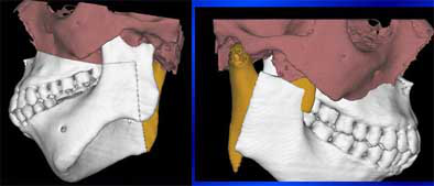
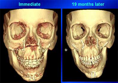
Above and below: 17-year-old girl with vertical maxillary excess, maxillary and mandibular asymmetry, and a hypoplastic condyle. Multiplanar maxillary-mandibular segmentation technique with SSD 3D reconstruction was used for presurgical planning. Anterior nasal spine, left malar prominence, and finally the left mandibular condyle were tested as trial pivot points to produce the best result with the least amount of surgery. Mandibular asymmetry had re-emerged 19 months postsurgery, however. Currently 3D CT reconstructions are being compared to isolate the cause of the relapse.
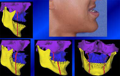
Another patient recently presented with vertical maxillary excess, mandibular hypoplasia, labial dental protrusion, and severe periodontitis, Bradrick said. She wanted dentures, "but you can't even make dentures with this (protrusion) in the middle," he said. Volumetric CT images were acquired to plan a multipiece maxillary osteotomy.
"We did all this (planning) in virtual space," Bradrick said. We select a pivot point and axis of rotation, spin the (maxilla) back, then select a point of rotation in the condyle region, and ... fix it so there's no vertical excess anymore, measure it and do the osteotomy, and see if you end up where you want to be." The teeth were taken out last and replaced with dentures.
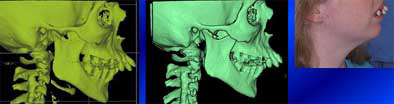
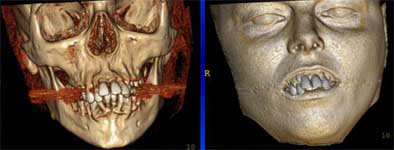
Above and below: Patient with vertical maxillary excess, mandibular hypoplasia, labial dental protrusion, and severe periodontitis underwent presurgical planning with CT volume reconstructions. Anterior segmental osteotomy was used to retract the maxilla by 10 mm. Then the teeth were removed, allowing the patient to have new dentures fitted.
Maxillary advancement
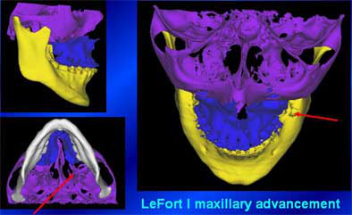
Maxillary advancement surgery is an important tool for normalizing facial contours and correcting dental occlusions. CT with volumetric reconstruction can replace the extensive photographic and modeling analysis used to plan bone-grafting techniques used to advance, retract, or tilt the maxilla in surgery. Horizontal osteotomy genioplasty, meticulously planned with CT reconstructions, can give the chin a more normal appearance than is possible with traditional alloplastic implants.
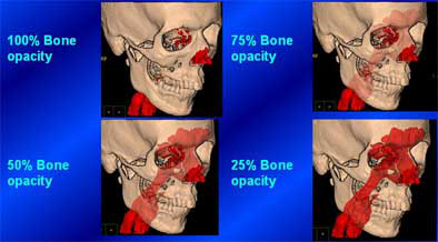
Markers (red) can be added to CT reconstructions to plan the adjustments in maxillary advancement surgery, replacing plaster modeling. Segmentation can also be performed with vectors and planes, which are especially helpful in planning genioplasty. The surgeon can move and manipulate the planes until the result is right.
Dental implants
At least three vendors of dental implant reconstruction software had booths at the AAOMS meeting, Bradrick said, and all of the programs seem to operate similarly. These applications allow the oral surgeon to simulate and draw in the alveolar canal, "inserting" implants to evaluate their relation to the canal, and measure the distance between the implant and the inferior alveolar nerve (IAN) that must remain intact.
Two recent dental implant cases resulted from the use of a published but ill-conceived anesthetic technique for dental implant surgery, Bradrick said.
"To put in a mandibular implant, some surgeons aren't using the traditional inferior alveolar nerve block. Surgeons are just infiltrating the area of implant placement with local anesthetic," he said, describing the primitive technique. "Supposedly when you're getting close to an unanesthetized inferior alveolar nerve, the patient will complain of pain and you stop."
But the pain warning didn't work in two recent cases, resulting in violation of the inferior alveolar canal with accompanying loss of sensation in the jaw. Fortunately, he said, the damage in the second case was mostly reversed.
"This was a relatively new case, only about a day old. They took out the implant real quick, and she got about 90% of her sensation back," he said. "You can almost see the threads on that implant."
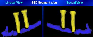
Above and below: Violation of the inferior alveolar canal in two dental implant cases, seen in reconstructed CT images. The mandibular loss of sensation was reversed in the second case (below), thanks to quick removal of the implant.
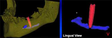
Few oral and maxillofacial surgeons are actually using CT reconstruction software at this point, he said, but the techniques will certainly gain more followers as CT scanners and better software become more widely available. CT with 3D reconstructions offers significant advantages in planning for trauma surgery, cranioplasty, orthognathic surgery, dental implants, and a number of other applications.
"Conebeam office-based CT units with multiplanar and 3D reconstruction software ... are now available if you want to spend $300,000," he said.
More than a decade ago, Fishman et al showed that 3D reconstructions CT altered the management of 20% to 30% of musculoskeletal patients, concluding that surgeons prefer 3D images for planning and patient management, Bradrick said (Radiology, November 1991, Vol. 181:2, pp. 321-327). In his practice, 3D volume reconstruction software has proved to be of great value in surgical planning -- and as an educational tool for patients.
"There are so many versatile things these programs can do, you're limited only by your imagination," Bradrick said.
By Eric Barnes
AuntMinnie.com staff writer
November 22, 2004
Copyright © 2004 AuntMinnie.com

One-hour tooth implant may have problemsBy Reuters Health


One-hour tooth implant may have problemsBy Reuters Health

July 18, 2007 -- STOCKHOLM (Reuters) Feb 19 Swiss-Swedish Nobel Biocare's Teeth-in-an-Hour implant system may carry more risk of complications than conventional methods, an abstract of a study by Sweden's Karolinska Institute shows.

The study examined 31 patients treated using the method, which uses computer modeling to reduce implant installation time. Complications such as the loss of fixtures and the need for adjustments occurred for nearly a third of the patients.

Nobel Biocare could not immediately be reached for comment.

Nine percent of the 175 fixtures examined--the titanium screws which are fastened to the bone--were lost, said Björn Klinge, professor of periodontology at the Karolinska Institute and one of the study's authors.

This compared with a loss rate of roughly 2% to 3% using conventional and more time-consuming methods, he added.

"My assessment is that this is still at the trial stage and therefore can't be recommended for general use," Klinge told Reuters.

"Our data shows rather unequivocally that there is a lot of development left before one can say if this is a good or a bad product," he added.

The study, presented at a dental conference in Venice on February 17, has not yet been published in a scientific journal or peer-reviewed, but Klinge said an article was being prepared.

He also noted the method offered several benefits, including shortened treatment time and reduced or eliminated bleeding, swelling and pain.

"In its way, this is a fantastic method and I think that there may be a lot of positive things coming out of this, but it is obvious that there are a number of problems which are not solved with this method," he said.

"If one is to use it, one should be clear that the risk of failure is up to 30%, and that is exceptionally high."

The study looked at implants using the Teeth-in-an-Hour method, unveiled in 2003, between three months and three years after installation.

Klinge is part of a panel of experts that advised Sweden's Medical Products Agency on another Nobel Biocare product, NobelDirect, after two Swedish professors said the implant caused what they called unacceptable levels of jawbone loss.

In a report to the agency that hit Nobel's share price, the panel said NobelDirect could not be regarded as a finished product and should only be used with caution under controlled circumstances.

Nobel Biocare has consistently claimed there is no problem with NobelDirect.

(Additional reporting by Sven Nordenstam)

Karolinska Institute

Copyright © 2007 Reuters Limited. All rights reserved. Republication or redistribution of Reuters content, including by framing or similar means, is expressly prohibited without the prior written consent of Reuters. Reuters shall not be liable for any errors or delays in the content, or for any actions taken in reliance thereon. Reuters and the Reuters sphere logo are registered trademarks and trademarks of the Reuters group of companies around the world.

Hep B virus transmitted to dental patientsBy Reuters Health


Hep B virus transmitted to dental patientsBy Reuters Health


July 16, 2007 -- NEW YORK (Reuters Health), May 2 Even with strict enforcement of infection control, hepatitis B virus (HBV) was transmitted from one patient to another when they both underwent oral surgery in the same office, on the same day, public health officials report.

The index case was a 60-year-old woman with joint pain, swelling, and fatigue that began in February 2002. She had no traditional HBV risk factors, the investigators report in the May 1st issue of The Journal of Infectious Diseases, but she said she had oral surgery several months earlier.

A cross-match of the state department of health's hepatitis B registry found a "source" patient infected with HBV who had oral surgery a couple hours earlier that day. Three other patients were treated in the intervening period.

According to Dr. John T. Redd and associates, from the Centers for Disease Control and Prevention in Atlanta, the source patient had chronic hepatitis B with a high viral load. The source patient and the index patient both had the A/subtype adw2 genotype and DNA sequencing indicated that the HBV isolates from the two patients were identical in the region of DNA examined.

Surveillance at the surgeon's office showed that appropriate, standard precautions for preventing transmission of blood-borne pathogens were being followed.

Transmission of HBV in a dental setting is rare. Nevertheless, HBV can persist in dried blood on surfaces for 1 week or longer, and can also be present on surfaces that have no detectable blood.

Transmission of HBV is about 100-fold more efficient than transmission of HIV in healthcare settings, Dr. Ban Mishu Allos and Dr. William Schaffner note in their accompanying commentary. Infection control has nearly eliminated its nosocomial spread. Still, several similar cases have been reported in which the means of transmission could not be identified, they add.

This is "troubling because it suggests that there are aspects of transmission of bloodborne disease that remain poorly understood," according to an editorial commentary from Drs. Ban Mishu Allos and William Schaffner of the Vanderbilt University School of Medicine in Nashville, Tennessee.

Allos and Schaffner advocate universal HBV vaccination of all adults up to 40 years of age, as well as thorough investigations for non-traditional exposure sources when HBV, HIV, or hepatitis C is diagnosed in patients with no recognizable risk factors.

MacPractice teams with Suni Medical Imaging, National Dental


MacPractice teams with Suni Medical Imaging, National Dental

June 17, 2008 -- MacPractice, developer of MacPractice DDS software, and Suni Medical Imaging, a manufacturer of intraoral sensors for dentistry, are collaborating to develop MacSensor, a Macintosh-based application comprising a new SuniRay sensor and MacPractice DR (digital radiography) software.

MacPractice DDS is a Mac-based dental practice management and clinical software application that includes a Mac OS X migration path for DentalMac users.

The new SuniRay sensor uses the latest CMOS technology to deliver high image quality for maximum diagnostic capabilities.

"This is a sensor solution for which dentists who prefer to use a Mac have been waiting," said Mark Hollis, MacPractice president.

In related news, MacPractice and National Dental have released the first direct-to-USB intraoral camera for the Mac. The system combines the DocPort Macro Intraoral Camera and MacPractice DR software to give MacPractice owners the ability to add high-quality intraoral images via USB to their operatories.

ADA seeks clarification from FDA on amalgam comments


ADA seeks clarification from FDA on amalgam comments

June 16, 2008 -- The ADA is expressing concern about recent statements from the FDA suggesting that the agency has already decided to mandate some form of special labeling for dental amalgam as a caution to groups purportedly sensitive to mercury.

The FDA is currently in the process of reviewing public comment on amalgam and possibly considering reclassifying it for regulatory purposes following the settlement of a lawsuit with consumer advocacy groups that had sought to have the material banned completely.

According to a press release from the ADA, a recent Associated Press article attributed this statement to FDA Deputy Commissioner Randall Lutter, Ph.D.: "What this says is there's a clear intent on our part on labeling for sensitive subpopulations."

The ADA does not believe that such action is supported by the best scientific studies published on this topic, and intends to seek clarification from FDA, including any scientific support for the agency's recent statements.

"People depend on the FDA and other government health agencies to help protect their health. It's critically important that public health recommendations are based on sound scientific evidence," states ADA President Mark J. Feldman, D.M.D. "The ADA will continue to advocate for the best oral health of the public as part of the FDA regulatory process."

Presently, FDA has different classifications for encapsulated amalgam and its component parts, dental mercury and amalgam alloy. The FDA's proposed reclassification, which the ADA has supported since 2002, would place encapsulated amalgam and its components under one classification.

Based on extensive studies and scientific reviews of dental amalgam by government and independent organizations worldwide, the ADA believes that dental amalgam remains a safe, affordable, and durable cavity filling choice for dental patients.

CAD/CAM competitor claims advantages over Cerec


CAD/CAM competitor claims advantages over Cerec

June 16, 2008 -- Imagine shopping for a car if the only make was Ford. Or dining out if the only place to eat was Sizzler. For 21 years, that has been the situation dentists faced if they wanted to make crowns and other restorations in their own offices.

This year, the scenario changed.

After years of advance publicity, Dallas company D4D Technologies launched its E4D CAD/CAM system in the first-ever direct challenge to Sirona Dental Systems' Cerec system. The newcomer, while slightly more expensive, offers innovations aimed at tempting dentists away from the more established competitor.

Most notably, dentists using E4D won't need to dust their patients' teeth with titanium powder before making the images needed to design restorations.

But analysts say the newcomer isn't likely to hurt Cerec -- at least in the short term. "Its commercial availability will stimulate adoption of chairside CAD/CAM technology in the U.S.," wrote Jon Wood, a stock analyst from Bank of America.

He and other observers say that the availability of the new system will prod buyers to finally get out their wallets. Many have been waiting to make a purchase until they could compare the two machines. Currently only about 8% of dentists use CAD/CAM in their offices.

And when DrBicuspid.com caught up with Sirona Marketing Coordinator Gabe Foster at the California Dental Association spring meeting, he said his company welcomes the rival. "We think that competition is great for the market," he said. "It pushes us to make sure our product is as good as it can be."

No studies offering a head-to-head test of which machine is faster or more reliable have so far been published. True comparisons may take years as researchers document how well restorations made on the two machines last.

But D4D is working hard to nudge the older technology aside. "We think we got it much better," said Mark Ebersold, director of dental affairs at D4D, as he showed DrBicuspid.com around the company's headquarters in suburban Dallas. "The Cerec is a great machine. It certainly works. If it was not for their effort, we would not be here. We believe we have taken it to a whole new level."

The machines look similar. For both, you sit at a console mounted on a cart. A computer is attached to a camera wand, monitor, keyboard, and mouse. You position the wand in the patient's mouth and take an image of the tooth to be restored. The image appears on the monitor. You use the mouse and keyboard to manipulate the image in three dimensions, as you design the restoration you want. Both machines do inlays, onlays, crowns, and veneers.

You then load the milling machine with the kind of material you want -- generally one of several types of ceramic. You press a button and the milling machine -- which can be in the same room or at some distance -- uses two opposing diamonds to cut the block into the desired shape.

Key differences

Ebersold stresses the differences in making images. While Cerec bounces infrared light off the tooth, E4D uses a laser. The wands are different shapes, and both companies claim that theirs is easier to get into the patient's mouth.

There is one significant difference, however: the Cerec can't make an image of the tooth surfaces without titanium dioxide being applied to the tooth first. "[You] have to maintain that dry and take a picture, and then clean up afterward," Ebersold said. "If you think about snow drifts, you get an idea of the detail you lose."

When pressed, Ebersold admits that in some cases -- he estimates less than 5% -- the enamel on a patient's tooth is so translucent that the E4D laser won't reflect sufficiently. He argues that this won't happen if dentists prepare the tooth as directed. But when it does, he says, dentists may either apply Accent, a reflective solution, or scan the tooth from other perspectives.

Another difference is that the Cerec scans just once and always from an occlusal perspective -- the equivalent of taking a single exposure. The E4D, on the other hand, takes images from several perspectives, including buccal and lingual.

Ebersold argues that this allows the E4D to obtain more detail. Foster responds that a single scan has more integrity. "When you're stitching images together, you have a high probability of inaccuracy," he said.

The two systems differ in the milling process as well. The Cerec computer has to keep communicating with the mill until the restoration is ready. The E4D sends instructions to a job server that feeds the information to the mill, freeing the digital cart for another job.

On the other hand, Cerec boasts faster milling speed. Though the time varies depending on the job, the latest Cerec model -- the MCXL -- ranges from four to 10 minutes per restoration, while the E4D "averages" 10 to 15 minutes, according to Ebersold. The E4D mill is also chunkier, weighing in at 250 lb compared to only 70 lb for the Cerec.

Foster is quick to point out another key advantage of Cerec: some 15,000 dentists already own one, so its strengths and weaknesses are pretty well known. If you buy an E4D, you'll be something of a pioneer. Just to look at one, you pretty much have to go to a trade show or fly to Dallas.

"We back up our claims with clinical results," said Foster, who claims a 95% success rate for Cerec restorations. Search Medline under "Cerec" and you'll get 359 hits. Try "E4D" and you'll get none.

There's no real price advantage for the new product either. The E4D runs $116,500. The top-of-the-line Cerec will set you back $109,995, but you can also get the older model with a mill time of eight to 17 minutes for $82,995, or a refurbished unit for $69,995.

Still, some pretty big names are backing D4D in its venture. Partners include Henry Schein (the distributor), 3M, and Ivoclar. Cerec is also associated with heavy hitters; it is distributed by Patterson.

So which machine will triumph? Perhaps neither one. If the market expands as expected, there will be plenty of room for both Ford and Toyota.

Beyond x-rays: Part I -- Do optical caries detection systems really work?


Beyond x-rays: Part I -- Do optical caries detection systems really work?

June 16, 2008 -- Imagine being able to find -- and perhaps even reverse -- a carious lesion before it's detectable on an x-ray. Imagine being able to "see" whether demineralization has begun and how advanced it might be.

X-rays and visual examination have long been the dentists' diagnostic methods of choice. But x-rays routinely miss 70% to 80% of occlusal caries. And even the most well-trained eye cannot see what havoc bacteria may be wreaking beneath the tooth's surface. Nor can it determine the degree of demineralization that may be taking place.

A number of products now offer dentists the ability to do just this, and in some cases even more. The key is light -- or, more precisely, photons. While these optical detection systems are not intended to replace visual examinations or radiography, they do provide additional information about what is going on in a tooth at the microscopic level. However, even for those products already on the market, the jury is still out on how useful they are beyond serving as an adjunct to x-rays and visual exams.

One of the first optical devices developed specifically for caries detection was the DIFOTI (digital imaging fiber-optic trans-illumination) system, developed and marketed by Electro-Optical Sciences. Introduced in 2001, it is the only instrument of its kind to be approved by the FDA for the detection of incipient, frank, and recurrent caries. A single fiber-optic cable in the patented mouthpiece delivers visible light to a tooth's smooth surfaces. As the light travels through layers of enamel and dentin, it scatters in all directions toward the nonilluminated surfaces. The light is then directed through the mouthpiece to a CCD camera in the handpiece. The camera digitally images the light emerging from the various surfaces of the teeth. These images are displayed on a computer monitor in real-time and stored on the hard drive.

A number of clinical studies involving DIFOTI have been conducted over the years, and the findings consistently support the viability of this device for imaging cracks and detecting some early caries. For example, a 2005 study that looked at the DIFOTI system for evaluating early approximal lesions concluded that although the device was not able to measure the depth of a lesion in any of the samples, it was able to show surface changes associated with early demineralization as early as two weeks (Journal of the American Dental Association, December 2005, Vol. 136:12, pp. 1682-1687).

DIFOTI initially sold for about $6,500. But in 2006 the company signed an exclusive licensing agreement with KaVo for all marketing, sales, service, and further development of the system. At this point, it is not clear what has happened to the product; there is still a DIFOTI Web site (www.difoti.com), but if you call the sales/service number on the site, you will be told that Electro-Optical Sciences no longer has anything to do with the product and that you should contact KaVo Dental. Repeated queries to KaVo went unanswered.

Glowing bacteria

KaVo is better known for another optical caries detection device, the Diagnodent. The system, which was introduced in 2003 and currently sells for $3,150, uses a red diode laser to measure the amount of fluorescing bacteria in a suspect area. As the laser beam passes over the tooth, the system "beeps" if any fluorescence is detected. A numerical value between 1 and 100 is then displayed, indicating the degree of decay being detected.

At the Diagnodent wavelength (655 nm), clean, healthy tooth structures exhibit little to no fluorescence. A reading between 15 and 30 may require a preventive fluoride prescription to encourage remineralization or restorative care, depending on the caries risk assessment. Numbers greater than 30 indicate the probability of advanced disease and the need for operative treatment.

While numerous clinical studies have demonstrated the viability of Diagnodent for enhancing caries detection (the KaVo Web site lists 27 articles from scientific journals, dating back to 1998) and the company currently claims that the Diagnodent system is being used in 20% to 25% of dental offices, its utility is limited.

According to a 2006 study (Journal of Clinical Dentistry, 2006, Vol. 17:3, pp. 53-58), "While the Diagnodent is an important diagnostic device, particularly for diagnosing hidden carious lesions in the dentine layer, it should be used along with visual examination in clinical practice, as [it] is not superior to visual examination in detecting early stages of carious lesions."

"We use it in our office, and the literature supports that it is a nice adjunct, but it should not be used by itself as a way to detect caries," said Edwin Parks, D.M.D., M.S., director of dental radiology at Indiana University and a member of the DrBicuspid.com advisory board. "Specificity is an issue with the Diagnodent. There are too many false positives that can come from the depth of the fissure or from staining. Threshold is a big issue also. At what point does a reading indicate a clinical issue, and what value indicates a carious lesion versus a deep pit?"

Another commercially available caries detection system uses light-emitting diodes rather than laser to detect decalcification and occlusal and interproximal caries. The D-Carie system was developed by Neks Technologies, which was acquired by Dentsply in 2007. D-Carie gained FDA clearance that same year and sells for $3,100, now under the name Midwest Caries I.D.

Like Diagnodent, the pen-sized, cordless device provides both an audible signal and visual cue (green light turns to red) when fluorescence from decay is detected. According to a study performed at the University of Montreal and presented at the 2006 International Association of Dental Research meeting, a comparison of caries detection using D-Carie and Diagnodent on 39 teeth found that sensitivity was 91% for D-Carie compared to 93% for Diagnodent, while specificity was 60% for D-Carie compared to 26% for Diagnodent.

"The D-Carie, which uses both IR [infrared] and light fluorescence, is better than Diagnodent for caries detection, but it still focuses on cavitated lesions," said George Stookey, Ph.D., distinguished professor emeritus at Indiana University and president and CEO of Therametric Technologies.

More useful would be the ability to measure early demineralization in tooth enamel, a precursor to decay, Stookey added. Therametric is working to commercialize a technique called quantitative light fluorescence (QLF) that can do just that.